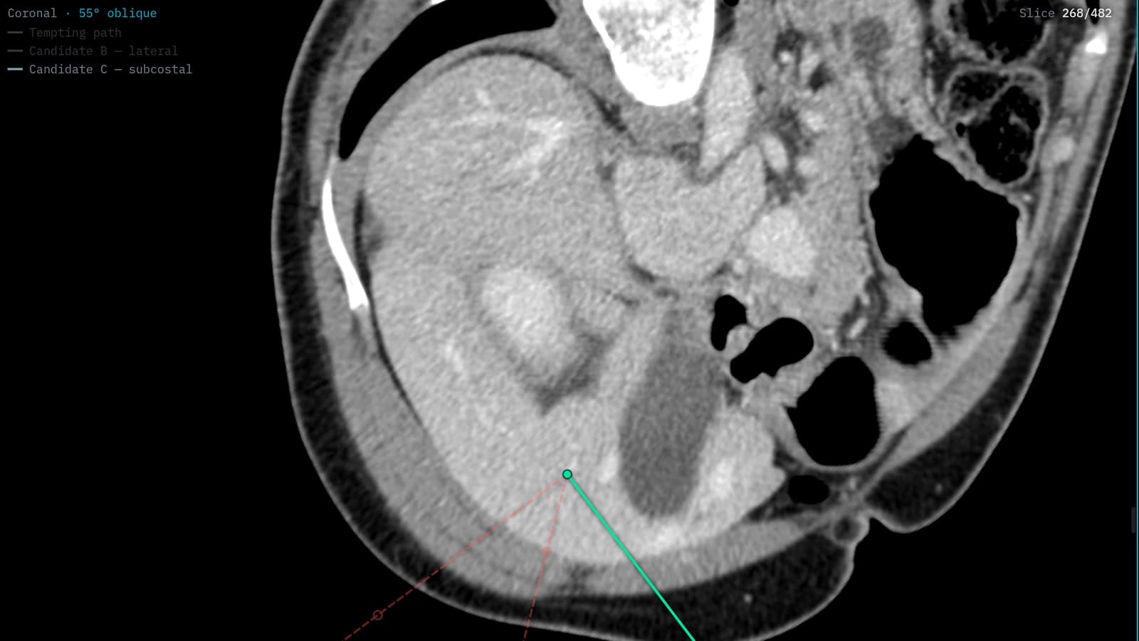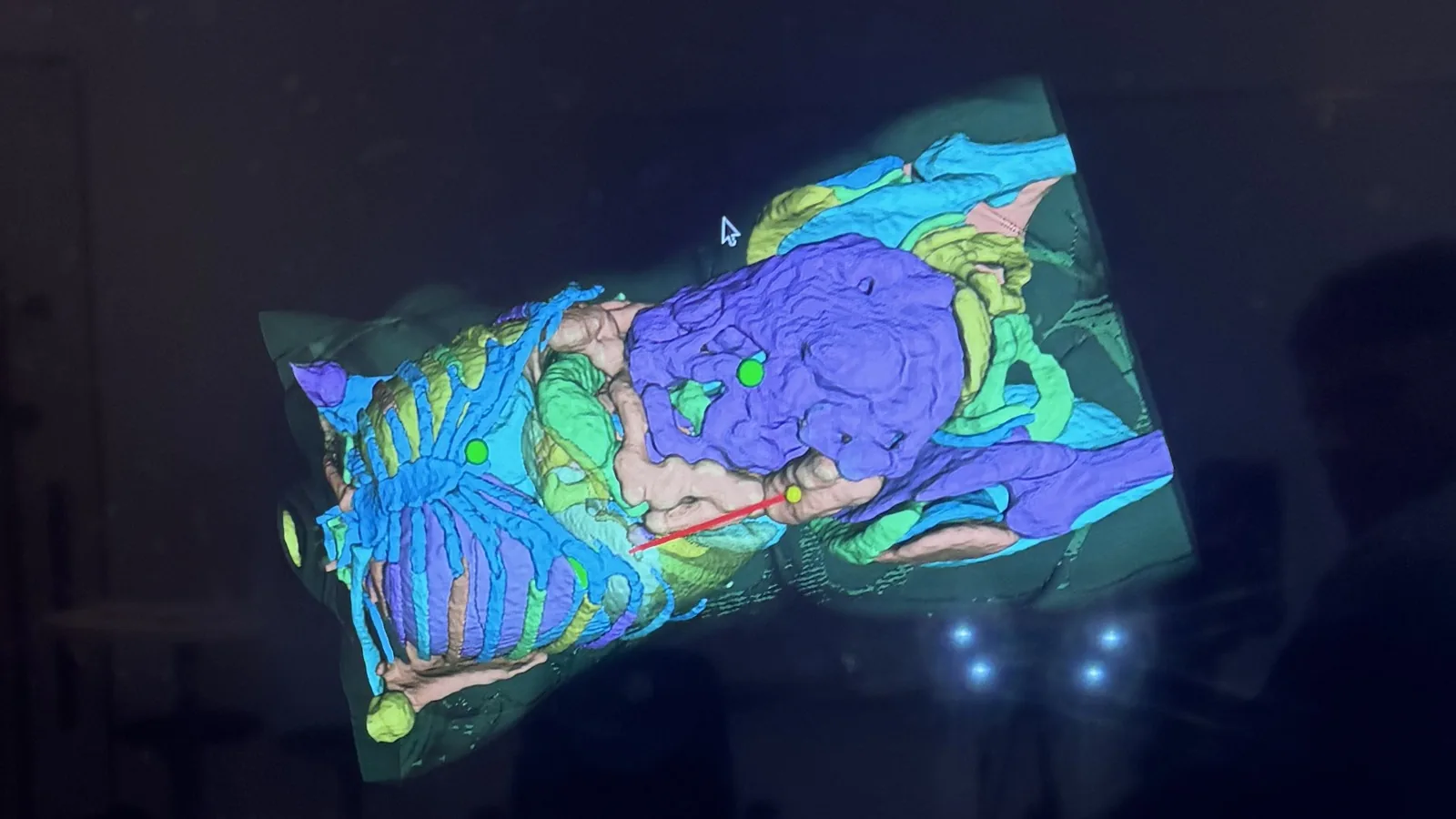
Percisio Navigation guides a needle to a target planned on the patient's CT. It is built for the everyday percutaneous procedures — biopsy, drainage, infiltration and puncture. A camera unit on a tripod registers the CT to the patient without anything attached to the body, tracks the needle in the operator's hand, and shows at every moment where the needle is against the planned trajectory.
We call it a stereotactic navigation system. Few words in image-guided intervention have a definition as precise, or as old, and few are used as loosely: in product copy, stereotactic usually means "very accurate". So this article does two things. It shows how Percisio Navigation works, step by step, and it checks each step against what the stereotactic method actually requires — including where the abdomen forces caveats that a skull never did. Every claim about stereotaxy itself is referenced, and the sources are listed at the end.
What "stereotactic" requires
In the literature, stereotactic means something narrower and more useful than "accurate": a method for reaching a point inside the body by its coordinates. The word joins two Greek roots: stereos, a solid — hence three-dimensional — and taxis, from tattein, to position (Grunert et al., 2015). The US National Library of Medicine still files the method under its older spelling, stereotaxic techniques: "techniques used mostly during brain surgery which use a system of three-dimensional coordinates to locate the site to be operated on" (MeSH D013238). The phrase that matters is a system of three-dimensional coordinates. Nothing in it requires a brain, or a frame.
There is no single checklist in the literature that every author signs up to. But take away the neurosurgical setting, and what the definitions and reviews of image-guided intervention share can be laid out in five parts. This is the working definition we use in this article:
- A three-dimensional image of the patient — the map in which the target is visible.
- A coordinate system tied to the patient, so that a point in the image and a point in the body can be the same point.
- Registration — the transformation that maps image coordinates onto the patient's physical coordinates.
- A plan expressed in those coordinates — a target and an entry point, which together define a trajectory.
- An instrument whose position is known in the same coordinates, either constrained by a frame or tracked, so that its deviation from the plan can be measured while it advances.
Judged this way, what makes a system stereotactic is the method, not a frame, an arm or a particular tracking technology. A system that shows a detailed 3D model but cannot place the instrument in that model's coordinates is visualisation. A system that follows the needle on live 2D images alone is image guidance. Both are useful; neither is stereotaxy. And having the five parts is not enough on its own: the link between image, patient and instrument has to be established, and kept, accurately enough for the procedure at hand.
Percisio Navigation has all five parts. The rest of this article shows how, and where keeping that link accurate is hardest.
How Percisio Navigation works, step by step
Each step of the method has a counterpart in the Percisio workflow.
The image. Navigation starts from the patient's CT. From it, Percisio builds what we call the hologram: a 3D model in which the skin, bones and organs are segmented automatically and the target is identified.
Coordinates and registration. This is the step that earns the word stereotactic, and the one where Percisio differs most from earlier systems. There is no frame and no reference array fixed to the patient. Percisio's camera unit estimates the patient's pose from the body itself — markerless body tracking, using computer vision and depth information — and registers the CT-derived model to it. Once the two are aligned, a point in the CT and a point in the patient correspond, within the error of the registration.
The cameras see the skin, not the organs. Registering to the body surface therefore rests on an assumption: that the organs sit where the CT showed them relative to that surface. Patient movement and breathing break that assumption, and the next section covers how Percisio deals with them. The accuracy that matters is measured at the target, not at the skin.
The plan. The operator defines the plan inside the 3D model: the target, the entry point and the needle path between them. During the procedure the display carries the planned path, the entry point and the remaining distance to the lesion. Choosing that path well is a skill in its own right; we wrote about teaching it in Teach the Plan, Not Just the Puncture.
The tracked instrument. The needle carries a lightweight attachment module that the cameras track in real time, so the needle's position and direction are known in the same coordinates as the plan. Guidance is the difference between the two: where the needle is, against where the plan says it should be.
Most navigation systems track instruments either optically, with cameras that follow marker arrays, or electromagnetically, with a field generator (Mezger et al., 2013). Each has known constraints: optical systems need a clear line of sight to their markers, and electromagnetic tracking, which avoids that constraint, brings error sources of its own, including distortion from metal in the field (Franz et al., 2014). Percisio uses camera-based computer vision instead; our technical presentation, embedded below, walks through that choice. The tracking technology changes; the stereotactic method does not.
Where the abdomen is harder than the skull
Stereotaxy depends on maintaining an accurate relationship between the target, the image and the instrument. In the skull, rigid fixation can make that relationship relatively stable. In the abdomen, breathing and tissue deformation make it harder to maintain, and a navigation system must account for those limitations.
Breathing moves the target. "Hepatic motion and deformation due to respiration remain obstacles to applying stereotactic localization techniques to the liver" (Clifford et al., 2002). In a classic measurement, the liver moved about 2.5 cm craniocaudally in normal breathing and 5.5 cm in maximal breathing (Suramo et al., 1984) — an order of magnitude more than the few millimetres a needle procedure can tolerate. A plan made on a CT acquired at one point in the breathing cycle holds only at the same point in the cycle. The Innsbruck group of Reto Bale and Gerlig Widmann, whose stereotactic liver work is described below, controls this under general anaesthesia by disconnecting the endotracheal tube during imaging and needle placement (Widmann et al., 2010).
Percisio does not try to predict where the organs have moved. It reports, in real time, how well the patient still matches the CT: the skin surface from the CT is compared with the surface the cameras see, and the operator is shown how well the two agree. When the patient breathes or moves away from the position of the CT, the match drops, and the operator pauses the gesture until it recovers. The Monitoring Breathing demonstration shows this in use.
That is monitoring, not motion compensation, and the distinction matters. The match is measured on the skin, not at the target. A good match indicates that the external surface resembles the CT-derived surface; it does not prove that internal organs have returned to their original positions. A poor match is a clear warning that the plan may no longer be reliable. The clinical significance of the surface-match score therefore depends on how well it correlates with target-position accuracy under the intended operating conditions.
Registration error is not target error. How well the registration points line up says little about the error at the target you actually care about (Fitzpatrick et al., 1998; Fitzpatrick, 2009). The honest figure for a navigation system is measured at the target, along the instrument. Ours is a mean guidance error of 2 mm or less on the needle trajectory, measured on a test bench and an anatomical phantom. It is a bench and phantom figure, not a clinical one, and we present it as such.
Some targets are smaller than the error budget. Percisio uses depth information and the size of the target volume to decide whether guidance can be offered, and only validates actions inside its precision range. When a target is too small or too deep for that range, Percisio guides the initial trajectory and brings the needle as close as it safely can; the operator completes the final step under CT control. Stereotaxy does not replace control imaging. It reduces how much of it the approach needs.
Stereotactic navigation today: what CAS-One IR established
Among stereotactic systems for percutaneous abdominal work, CAS-One IR, made by CAScination in Bern, has one of the largest bodies of published evidence. Its US clearance describes it, in so many words, as "a user controlled, stereotactic accessory intended to assist in planning, navigation and manual advancement of one or more instruments … during Computed Tomography (CT) guided procedures" (FDA 510(k) K261225). That is the method described above, in regulatory language.
Several of the key clinical series in stereotactic liver ablation were gathered with it. Over 174 ablations of hepatocellular carcinoma in Bern, local tumour progression was 6.3% (Lachenmayer et al., 2019). A prospective registry at MD Anderson reported 7.4% local tumour progression at 12 months (Paolucci et al., 2026). More broadly, a retrospective two-centre study of hepatocellular carcinoma found incomplete ablation in 4.7% of lesions treated with CT-guided stereotactic navigation against 20.5% with ultrasound guidance — and 7.5% against 30.4% in the hard-to-reach segments II, IVa, VII and VIII (Perlberger et al., 2025). A meta-analysis across stereotactic and robotic systems put the weighted average lateral targeting error at 3.7 mm (Tinguely et al., 2021).
Taken together, that is strong evidence that stereotaxy works below the diaphragm. It also shows what it has required so far:
- A cart. CAS-One IR is a mobile platform 2 m tall, weighing 85 kg (K261225 summary).
- Fiducials on the patient. Registration relies on optical markers attached to the skin and captured in the planning CT (Tinguely et al., 2020).
- A mechanical arm, in the standard configuration. The needle is aligned through an aiming device on a multi-axis mechanical arm, then advanced by hand. A tracked marker clamped directly to a rigid needle is offered as an alternative (K152473 summary).
- General anaesthesia. Published workflows control breathing with high-frequency jet ventilation or breath holds under general anaesthesia (Lachenmayer et al., 2019; Paolucci et al., 2026).
For liver tumour ablation, the additional equipment and respiratory management used in published stereotactic workflows can be justified by the demands of the procedure. For routine biopsies, drainages and punctures, the practical requirements differ, and iterative CT-guided freehand placement remains one established approach (Arnolli et al., 2015). Percisio is designed to bring planned, tracked needle guidance to this broader range of procedures without requiring the same equipment configuration.
What Percisio changes
Percisio Navigation keeps the method and changes what it costs to apply it:
- Nothing attached to the patient. Registration is markerless, from the body the cameras see.
- No cart and no arm. The camera unit stands on a tripod and travels in a small bag. The needle stays in the operator's hand, carrying a lightweight module that the cameras track.
- Breathing monitored. A live score shows how well the patient's surface still matches the CT, so the operator advances the needle when it does and pauses when it does not.
- Built for the everyday procedures — biopsy, drainage, infiltration and puncture — so that stereotactic guidance is not reserved for the cases that justify a general anaesthetic.
What we will not do is put our accuracy figure next to theirs. The clinical figures above are measured in patients, often with different metrics from one study to the next. Our mean guidance error of 2 mm or less is measured on a bench and an anatomical phantom. Setting them side by side would flatter us and mean nothing. That comparison has to be made in patients.
Where the method comes from: the frame was never the point
Markerless registration and camera tracking are only the latest change of hardware in a method more than a century old — older than the medical images it now depends on. Each generation replaced the hardware while keeping the steps.
1908 — coordinates without images. Victor Horsley and Robert Clarke built an apparatus to place electrodes in the cerebellum of animals using Cartesian coordinates referenced to landmarks on the skull (Horsley & Clarke, 1908). The target's position came from an atlas, not from an image of the individual animal.
1947 — the patient's own image. Ernest Spiegel and Henry Wycis brought the method to humans and made the decisive change: coordinates were referenced to internal landmarks visible on the patient's own radiographs rather than to the outside of the skull (Spiegel et al., 1947). The target was now located in an image of this patient.
1949 — the arc. Lars Leksell's frame was built on the arc-centred principle: the target sits at the centre of the arc, so a probe advanced along any trajectory within the arc's range arrives at the same point (Leksell, 1949; Zrinzo, 2012). Once the target was fixed, the trajectory became a choice.
1979 — CT. Russell Brown's N-shaped localiser let the CT scanner's own slices encode frame coordinates, so targets could be located directly in a three-dimensional CT dataset (Brown, 1979). CT and stereotaxy have been inseparable since.
1986–1987 — frameless. David Roberts and colleagues tracked an operating microscope with a sonic digitiser and registered it to CT (Roberts et al., 1986). Eiju Watanabe and colleagues' Neuronavigator used an articulated arm to report a pointer's position in CT coordinates (Watanabe et al., 1987). The frame had been doing two jobs — defining coordinates and holding the instrument. Registration and tracking took both over, and both papers still called the method stereotaxic.
2000s — the abdomen. Optical tracking carried the same steps to the liver. The Innsbruck group of Bale and Widmann describes its CT-planned, optically navigated needle placement as frameless stereotaxy: "the spatial information gained from 3D imaging is available in coordinates" (Bale et al., 2010). Across 177 liver lesions treated by stereotactic radiofrequency ablation, the group reported 95.5% primary technique effectiveness and no significant difference between operators (Widmann et al., 2012). The same method applied to liver biopsy gave a diagnostic sample in 45 of 46 cases (Schullian et al., 2011).
Across that century, what establishes the coordinates keeps changing — skull landmarks, radiographic landmarks, frame fiducials on CT, digitisers, optical trackers. The steps stay recognisably the same. Reviews of image-guided intervention describe the same core: imaging, registration and tracking (Peters, 2006). Percisio Navigation keeps that core and replaces the fiducials, the cart and the arm.
See it running

Percisio's technical choices compared with electromagnetic and infrared tracking, and how the system determines the patient's pose. More demonstrations — breathing, multi-camera setups, augmented reality — are in the video library.
We will demonstrate Percisio Navigation live at MEDICA 2026 in Düsseldorf, 16–19 November, at Hall 17, Stand B45. If you would rather see it on your own studies, book a demo.
References
- Arnolli MM, Hanumara NC, Franken M, Brouwer DM, Broeders IAMJ. An overview of systems for CT- and MRI-guided percutaneous needle placement in the thorax and abdomen. International Journal of Medical Robotics and Computer Assisted Surgery. 2015;11(4):458–475. doi:10.1002/rcs.1630
- Bale R, Widmann G, Stoffner R. Stereotaxy: breaking the limits of current radiofrequency ablation techniques. European Journal of Radiology. 2010;75(1):32–36. doi:10.1016/j.ejrad.2010.04.013
- Brown RA. A computerized tomography-computer graphics approach to stereotaxic localization. Journal of Neurosurgery. 1979;50(6):715–720. doi:10.3171/jns.1979.50.6.0715
- Clifford MA, Banovac F, Levy E, Cleary K. Assessment of hepatic motion secondary to respiration for computer assisted interventions. Computer Aided Surgery. 2002;7(5):291–299. doi:10.3109/10929080209146038
- Fitzpatrick JM. Fiducial registration error and target registration error are uncorrelated. Proceedings of SPIE 7261, Medical Imaging 2009: Visualization, Image-Guided Procedures, and Modeling. 2009:726102. doi:10.1117/12.813601
- Fitzpatrick JM, West JB, Maurer CR Jr. Predicting error in rigid-body point-based registration. IEEE Transactions on Medical Imaging. 1998;17(5):694–702. doi:10.1109/42.736021
- Franz AM, Haidegger T, Birkfellner W, Cleary K, Peters TM, Maier-Hein L. Electromagnetic tracking in medicine — a review of technology, validation, and applications. IEEE Transactions on Medical Imaging. 2014;33(8):1702–1725. doi:10.1109/TMI.2014.2321777
- Grunert P Sr, Keiner D, Oertel J. Remarks upon the term stereotaxy: a linguistic and historical note. Stereotactic and Functional Neurosurgery. 2015;93(1):42–49. doi:10.1159/000366490
- Horsley V, Clarke RH. The structure and functions of the cerebellum examined by a new method. Brain. 1908;31(1):45–124. doi:10.1093/brain/31.1.45
- Lachenmayer A, Tinguely P, Maurer MH, et al. Stereotactic image-guided microwave ablation of hepatocellular carcinoma using a computer-assisted navigation system. Liver International. 2019;39(10):1975–1985. doi:10.1111/liv.14187
- Leksell L. A stereotaxic apparatus for intracerebral surgery. Acta Chirurgica Scandinavica. 1949;99:229–233.
- Mezger U, Jendrewski C, Bartels M. Navigation in surgery. Langenbeck's Archives of Surgery. 2013;398(4):501–514. doi:10.1007/s00423-013-1059-4
- National Library of Medicine. Stereotaxic Techniques. MeSH descriptor D013238. meshb.nlm.nih.gov
- Paolucci I, Albuquerque J, Siddiqi NS, et al. Stereotactic percutaneous thermal ablation of primary and metastatic liver tumors: a prospective registry-based study. Journal of Vascular and Interventional Radiology. 2026;37(8):108855. doi:10.1016/j.jvir.2026.108855
- Perlberger G, Barialei I, Lalov L, et al. Ultrasound guidance versus CT guidance with stereotactic navigation in thermal ablation for hepatocellular carcinoma: a comparative retrospective analysis. European Journal of Surgical Oncology. 2025;51(9):110292. doi:10.1016/j.ejso.2025.110292
- Peters TM. Image-guidance for surgical procedures. Physics in Medicine and Biology. 2006;51(14):R505–R540. doi:10.1088/0031-9155/51/14/R01
- Roberts DW, Strohbehn JW, Hatch JF, Murray W, Kettenberger H. A frameless stereotaxic integration of computerized tomographic imaging and the operating microscope. Journal of Neurosurgery. 1986;65(4):545–549. doi:10.3171/jns.1986.65.4.0545
- Schullian P, Widmann G, Lang TB, Knoflach M, Bale R. Accuracy and diagnostic yield of CT-guided stereotactic liver biopsy of primary and secondary liver tumors. Computer Aided Surgery. 2011;16(4):181–187. doi:10.3109/10929088.2011.578367
- Spiegel EA, Wycis HT, Marks M, Lee AJ. Stereotaxic apparatus for operations on the human brain. Science. 1947;106(2754):349–350. doi:10.1126/science.106.2754.349
- Suramo I, Päivänsalo M, Myllylä V. Cranio-caudal movements of the liver, pancreas and kidneys in respiration. Acta Radiologica: Diagnosis. 1984;25(2):129–131. doi:10.1177/028418518402500208
- Tinguely P, Frehner L, Lachenmayer A, et al. Stereotactic image-guided microwave ablation for malignant liver tumors — a multivariable accuracy and efficacy analysis. Frontiers in Oncology. 2020;10:842. doi:10.3389/fonc.2020.00842
- Tinguely P, Paolucci I, Ruiter SJS, et al. Stereotactic and robotic minimally invasive thermal ablation of malignant liver tumors: a systematic review and meta-analysis. Frontiers in Oncology. 2021;11:713685. doi:10.3389/fonc.2021.713685
- US Food and Drug Administration. 510(k) premarket notification K152473: CAS-One IR (CAScination AG). 2016. accessdata.fda.gov
- US Food and Drug Administration. 510(k) premarket notification K261225: CAS-One IR (CAScination AG). 2026. accessdata.fda.gov
- Watanabe E, Watanabe T, Manaka S, Mayanagi Y, Takakura K. Three-dimensional digitizer (neuronavigator): new equipment for computed tomography-guided stereotaxic surgery. Surgical Neurology. 1987;27(6):543–547. doi:10.1016/0090-3019(87)90152-2
- Widmann G, Schullian P, Haidu M, Bale R. Stereotactic radiofrequency ablation (SRFA) of liver lesions: technique effectiveness, safety, and interoperator performance. CardioVascular and Interventional Radiology. 2012;35(3):570–580. doi:10.1007/s00270-011-0200-4
- Widmann G, Schullian P, Haidu M, Wiedermann FJ, Bale R. Respiratory motion control for stereotactic and robotic liver interventions. International Journal of Medical Robotics and Computer Assisted Surgery. 2010;6(3):343–349. doi:10.1002/rcs.343
- Zrinzo L. Pitfalls in precision stereotactic surgery. Surgical Neurology International. 2012;3(Suppl 1):S53–S61. doi:10.4103/2152-7806.91612
Accuracy figures for Percisio Navigation come from test-bench and anatomical-phantom measurements, not from clinical studies.
About Percisio
Percisio is a medical technology company developing AR-powered guidance for image-guided procedures. The platform enhances precision, reduces radiation exposure, and supports clinicians across interventional radiology and related specialties.
Get in touch